IMPORTANT SAFETY INFORMATION
WARNINGS AND PRECAUTIONS
Seizures: Seizures occurred in 37% of patients treated with GLIADEL Wafers for recurrent glioma in the recurrent high-grade glioma trial. New or worsening (treatment emergent) seizures occurred in 20% of patients; 54% of treatment emergent seizures occurred within the first 5 post-operative days. The median time to onset of the first new or worsened post-operative seizure was four days. Institute optimal anti-seizure therapy prior to surgery. Monitor patients for seizures postoperatively.
Intracranial Hypertension: Brain edema occurred in 23% of patients with newly diagnosed glioma treated with GLIADEL Wafers in the newly-diagnosed high-grade glioma trial. Additionally, one GLIADEL- treated patient experienced intracerebral mass effect unresponsive to corticosteroids which led to brain herniation. Monitor patients closely for intracranial hypertension related to brain edema, inflammation, or necrosis of the brain tissue surrounding the resection. In refractory cases, consider re-operation and removal of GLIADEL Wafers or Wafer remnants.
Impaired Neurosurgical Wound Healing: Impaired neurosurgical wound healing including wound dehiscence, delayed wound healing, and subdural, subgaleal, or wound effusions occur with GLIADEL Wafer treatment. In the newly-diagnosed high-grade glioma trial, 16% of GLIADEL Wafer-treated patients with newly diagnosed glioma experienced impaired intracranial wound healing and 5% had cerebrospinal fluid leaks. In the recurrent high-grade glioma trial, 14% of GLIADEL Wafer-treated patients with recurrent high-grade glioma experienced wound healing abnormalities. Monitor patients post-operatively for impaired neurosurgical wound healing.
Meningitis: Meningitis occurred in 4% of patients with recurrent glioma receiving GLIADEL Wafers in the recurrent high-grade glioma trial. Two cases of meningitis were bacterial; one patient required removal of the Wafers four days after implantation; the other developed meningitis following reoperation for recurrent tumor. One case was diagnosed as chemical meningitis and resolved following steroid treatment. In one case the cause was unspecified, but meningitis resolved following antibiotic treatment. Monitor postoperatively for signs of meningitis and central nervous system infection.
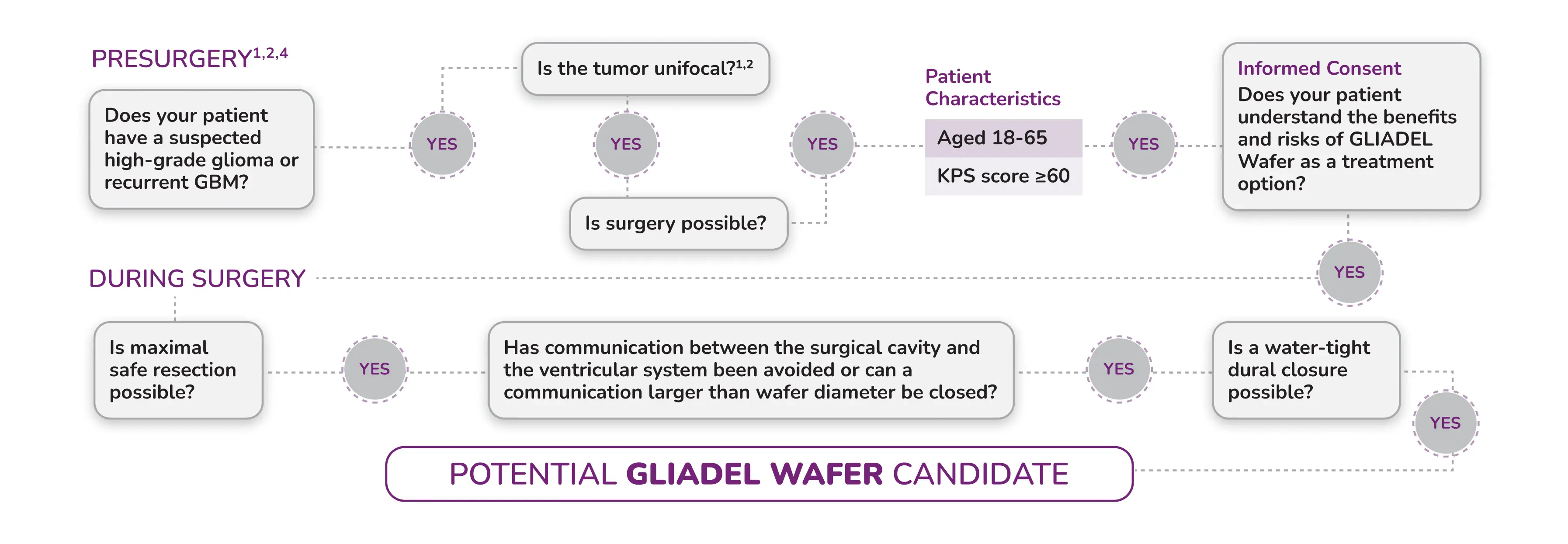
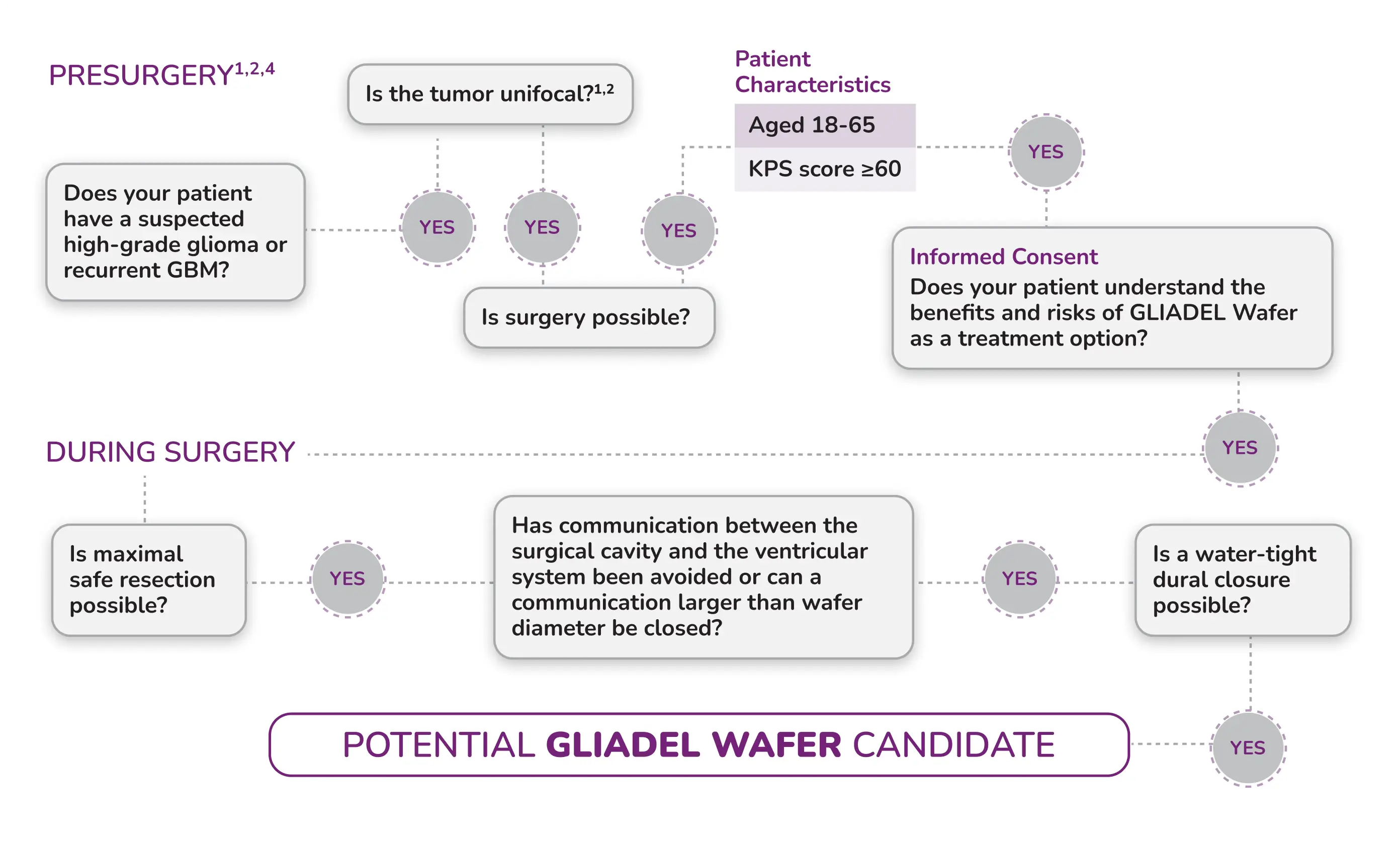
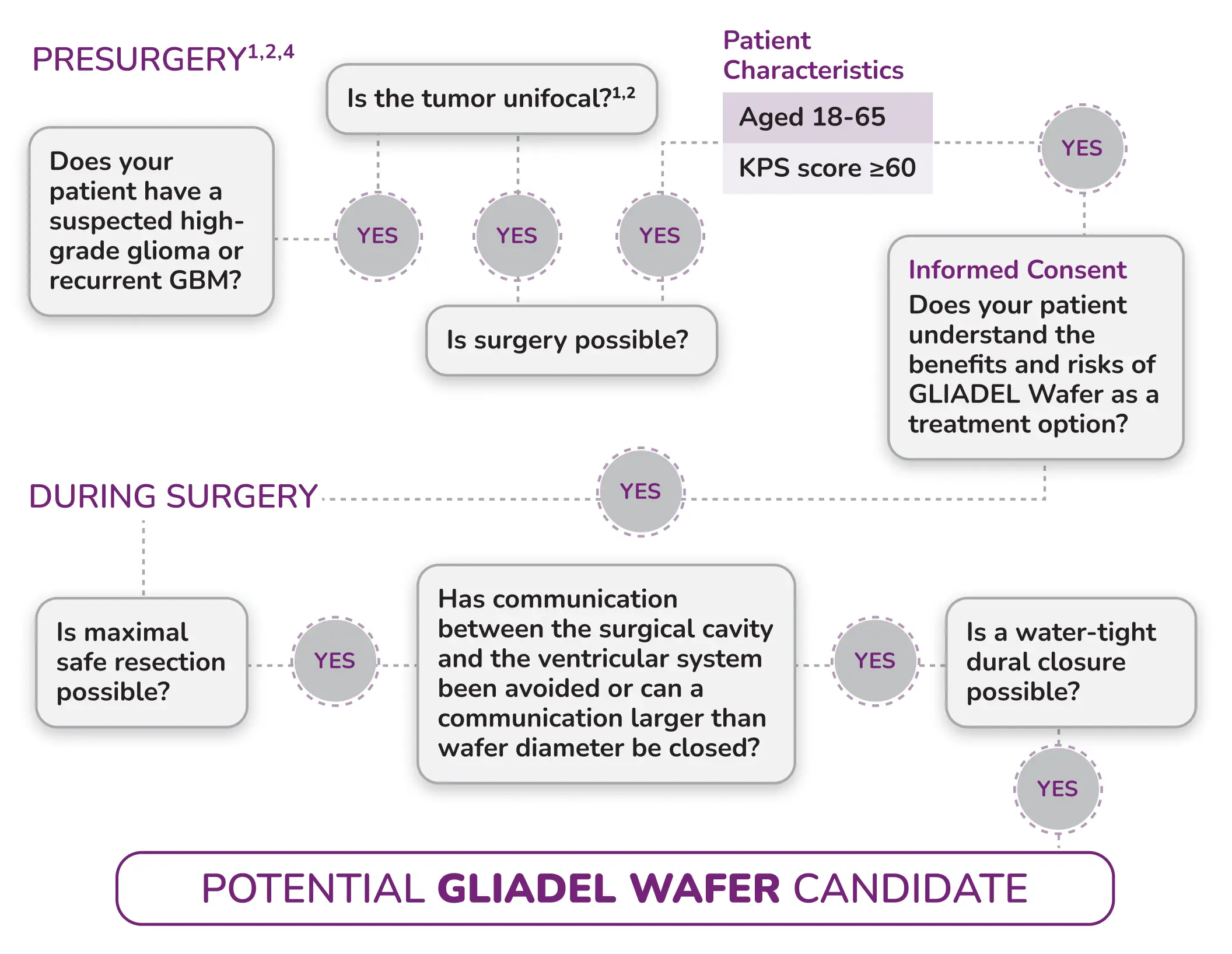
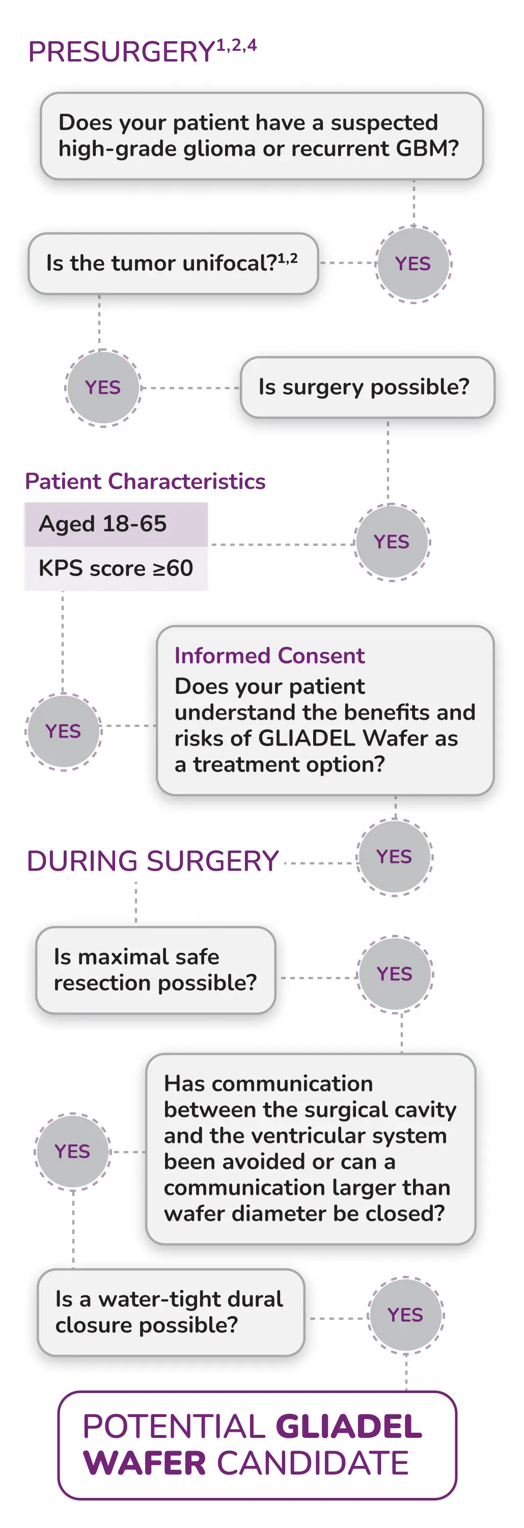
Wafer Migration: GLIADEL Wafer migration can occur. To reduce the risk of obstructive hydrocephalus due to wafer migration into the ventricular system, close any communication larger than the diameter of a Wafer between the surgical resection cavity and the ventricular system prior to Wafer implantation. Monitor patients for signs of obstructive hydrocephalus.
Embryo-Fetal Toxicity: GLIADEL Wafers can cause fetal harm when administered to a pregnant woman. Carmustine, the active component of GLIADEL Wafer, is embryotoxic and teratogenic in rats at exposures less than the exposure at the recommended human dose based on body surface area (BSA) and embryotoxic in rabbits at exposures similar to the exposure at the recommended human dose based on BSA. Advise patients of the potential risk to a fetus. Advise females of reproductive potential to use effective contraception for 6 months after implantation of GLIADEL Wafer. Advise males with female partners of reproductive potential to use effective contraception for 3 months following implantation of Gliadel Wafers.
ADVERSE REACTIONS
The most common adverse reactions in newly-diagnosed high-grade glioma patients (incidence >10% and between arm difference ≥4%) are cerebral edema, asthenia, nausea, vomiting, constipation, wound healing abnormalities and depression.
The most common adverse reactions in recurrent high-grade glioma patients (incidence >10% and between arm difference ≥4%) are urinary tract infection, wound healing abnormalities and fever.
These are not all the possible side effects of GLIADEL. Please see Full Prescribing Information for a full list.
The Important Safety Information does not include all the information needed to use GLIADEL safely and effectively. For additional safety information, please consult the full Prescribing Information for GLIADEL.
To report SUSPECTED ADVERSE REACTIONS, contact Azurity Pharmaceuticals, Inc. at 1-800-461-7449 or FDA at www.fda.gov/MedWatch or call 1-800-FDA-1088.
©2025 Azurity Pharmaceuticals, Inc.
PP-GLD-US- 0477